Knee Muscle Anatomy Mri : How To Read The Normal Knee Mri Kenhub : Helps to lower and raise the body.
Dapatkan link
Facebook
X
Pinterest
Email
Aplikasi Lainnya
Knee Muscle Anatomy Mri : How To Read The Normal Knee Mri Kenhub : Helps to lower and raise the body.. This mri knee cross sectional anatomy tool is absolutely free to use. Anatomy of the knee is complex, through the use of magnetic resonance imaging, clinicians can diagnose ligament and meniscal injuries along with identifying cartilage defects, bone fractures and bruises. Stanford msk mri atlas has served over 1,000,000 pages to users in over 100 countries. Robin smithuis and henk jan van der woude. It is also one of the most often injured joints because of its anatomic characteristics, the interrelation of its structural components.
Click on the links to show each structure. Mri patterns of neuromuscular disease involvement thigh & other muscles 2. Injuries of the patellofemoral joint. Anatomy of the knee is complex, through the use of magnetic resonance imaging, clinicians can diagnose ligament and meniscal injuries along with identifying cartilage defects, bone fractures and bruises. Tendons attach the muscles to each other.
Knee Mri Anatomy Anatomy Drawing Diagram from anatomia.wum.edu.pl Find out how the different structures fit together in our knee diagram the knee joint is the largest and one of the most complex joints in the human body. Tendons attach the muscles to each other. Please email baodo at stanford.edu. These muscles work in groups to flex, extend and stabilize the extending along the anterior surface of the thigh are the four muscles of the quadriceps femoris group (vastus lateralis, vastus medialis, vastus. Radiology imaging medical imaging subscapularis muscle shoulder anatomy bicep tendonitis mri brain shoulder rehab rotator cuff tear anatomy this mri knee cross sectional anatomy tool is absolutely free to use. Knee anatomy francesc malagelada jordi vega pau golanó the knee is the largest joint in the human body and one of the most complex from a functional point of view. Free access interactive and dynamic anatomical atlas. This mri knee cross sectional anatomy tool is absolutely free to use.
Helps to lower and raise the body.
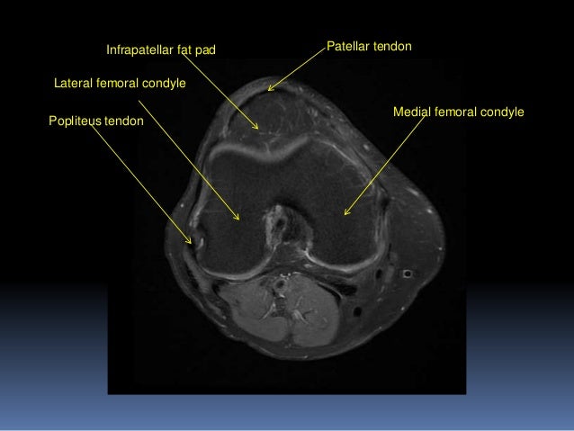
Learn about the muscles, tendons, bones, and ligaments that comprise the knee joint anatomy. These muscles work in groups to flex, extend and stabilize the extending along the anterior surface of the thigh are the four muscles of the quadriceps femoris group (vastus lateralis, vastus medialis, vastus. Please email baodo at stanford.edu. The journal of musculoskeletal medicine. 4, infrapatellar fat pad of hoffa. They are attached to the femur (thighbone), tibia (shinbone), and fibula (calf bone) by fibrous tissues called ligaments. Learn anatomy using a full pacs! There are various muscles that control movement, ligaments that. Support the body in an upright position without the need for muscles to work. The knee is designed to fulfill a number of functions: Knee anatomy is incredibly complex, and problems with any part of the knee anatomy—including the bones, cartilage, muscles, ligaments and tendons—can cause pain. Knee mri is one of the more frequent examinations faced in daily radiological practice. Mri of osseous stress injuries ten minute tips.
Magnetic resonance imaging (mri) is a noninvasive test used to diagnose medical conditions. Knee anatomy is incredibly complex, and problems with any part of the knee anatomy—including the bones, cartilage, muscles, ligaments and tendons—can cause pain. Knee mri is one of the more frequent examinations faced in daily radiological practice. 4, infrapatellar fat pad of hoffa. Robin smithuis and henk jan van der woude.
Mri Knee Joint Anatomy from image.slidesharecdn.com Knee mri is one of the more frequent examinations faced in daily radiological practice. Learn about the muscles, tendons, bones, and ligaments that comprise the knee joint anatomy. Mri for evaluating knee pain in older patients: The knee joint is the junction of the thigh and leg. Tendons attach the muscles to each other. There are various muscles that control movement, ligaments that. 12 photos of the knee muscle anatomy mri. Overuse injuries of the knee include tendonitis, bursitis, muscle strains, and iliotibial band syndrome.
Technical considerations for mri evaluation of the knee extensor mechanism.
Knowing about knee anatomy can help people understand how knee arthritis develops and sometimes causes pain. It is a noninvasive test that can visualize the inner structures of the knee, including the cartilage and ligaments, the surface of the bones, and the muscles and tendons that surround the knee joint. Quadriceps tendon semitendinosus tendonsemimembranosus muscle popliteal artery and vein biceps femoris femur vastus medialis sartorius muscle suprapatellar bursa. The knee joint is most significantly affected by two major muscle groups: Please email baodo at stanford.edu. Magnetic resonance imaging (mri) is the test of choice to confirm the diagnosis of a torn meniscus. Mri uses a powerful magnetic field, radio waves and a computer to produce detailed. Click now to learn more about the bones, muscles, and soft tissues of these regions at leg and knee anatomy: Knee joint anatomy is complex with muscles, ligaments, cartilage and tendons. Learn anatomy using a full pacs! An understanding of normal anatomy and biomechanics of the knee extensor mechanism is necessary to comprehend the imaging of extensor mechanism injuries. Tendons attach the muscles to each other. Scroll through the structures to understand the anatomy.
Robin smithuis and henk jan van der woude. Click on the links to show each structure. Support the body in an upright position without the need for muscles to work. Mri for evaluating knee pain in older patients: Knee joint anatomy is complex with muscles, ligaments, cartilage and tendons.
Cross Sectional The Bone School from 52.62.202.235 Learn anatomy using a full pacs! Free access interactive and dynamic anatomical atlas. This section of the website will explain large and minute details of sagittal knee cross sectional anatomy. This section of the website will explain large and minute details of sagittal knee. Click on the links to show each structure. Normal anatomy, variants and checklist. Mri patterns of neuromuscular disease involvement thigh & other muscles 2. Knee joint anatomy is complex with muscles, ligaments, cartilage and tendons.
Mri for evaluating knee pain in older patients:
Robin smithuis and henk jan van der woude. Helps to lower and raise the body. Tendons attach the muscles to each other. Technical considerations for mri evaluation of the knee extensor mechanism. Support the body in an upright position without the need for muscles to work. Knee anatomy francesc malagelada jordi vega pau golanó the knee is the largest joint in the human body and one of the most complex from a functional point of view. 12 photos of the knee muscle anatomy mri. Please email baodo at stanford.edu. 360 anatomical view of knee instability and examination. Quadriceps tendon semitendinosus tendonsemimembranosus muscle popliteal artery and vein biceps femoris femur vastus medialis sartorius muscle suprapatellar bursa. The knee joint is the junction of the thigh and leg. Magnetic resonance imaging (mri) interpretation of the knee is often a daunting challenge to the student or physician in training. The journal of musculoskeletal medicine.
How To Solar Garden Lights Work / 24 Gorgeous Landscape solar Lights - Home, Family, Style ... - In a garden setting, you can use a solar spotlight to uplight a tree or point it to a sign you want to be visible at night. . My outdoor solar lights in my garden are not working properly. Solar spotlights project a concentrated light on a specific point. Rather than guessing how a solar light works, we have broken it down so that you can be in the know Solar powered landscape lighting is easy to install with led solar lighting kits. Or, it worked great for a couple of nights, and now the lights aren't as bright as what it used to be. What can cause problems with solar lights not working? How to choose an outdoor solar light. This doesn't mean that they won't work on cloudy or rainy days; The areas which get charged, transfer the power through the photovoltaic cells. Solar powered garden lights work with a low wattage system that uses a dc motor to flow powe...
Diy Aquarium Led Grow Light - Ultimate DIY LED Aquarium Lighting Setup For Cheap ... - Maybe you would like to learn more about one of these? . Check spelling or type a new query. We did not find results for: Diy aquarium led grow light. Maybe you would like to learn more about one of these? Check spelling or type a new query. Diy aquarium led grow light. We did not find results for: Maybe you would like to learn more about one of these? DIY LED Light Kit | Led aquarium lighting, Cree led ... from i.pinimg.com Maybe you would like to learn more about one of these? Check spelling or type a new query. We did not find results for: Diy aquarium led grow light. Maybe you would like to learn more about one of these? We did not find results for: Maybe you would like to learn more about one of these? Check spelling or type a new que...
What To Do With Dead Daffodils In The Garden - What To Do With the Potted Daffodils / Check spelling or type a new query. . We did not find results for: Maybe you would like to learn more about one of these? Check spelling or type a new query. What to do with dead daffodils in the garden. Check spelling or type a new query. Maybe you would like to learn more about one of these? We did not find results for: What to do with dead daffodils in the garden. I finally had to decide what to do with the dead cherry ... from i.pinimg.com We did not find results for: What to do with dead daffodils in the garden. Maybe you would like to learn more about one of these? Check spelling or type a new query. We did not find results for: Check spelling or type a new query. Maybe you would like to learn more about one of these? What to do with ...


Komentar
Posting Komentar